Figure 2.
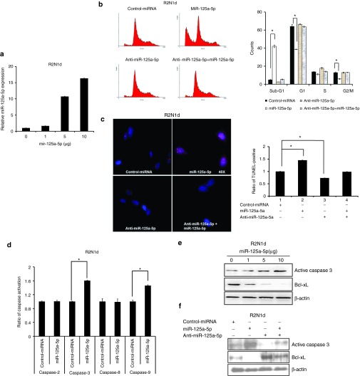
miR-125a-5p mediates the apoptosis signaling pathway. The control miRNA (5 μg/ml), miR-125a-5p (5 μg/ml), anti-miR-125a-5p (150 nmol/l), or anti-miR-125a-5p (150 nmol/l) + miR-125a-5p (5 μg/ml) were transfected into R2N1d cells and the cells assessed at 48 hours post-transfection. (a) MiR-125a-5p expression was analyzed by q-PCR with a dose-dependent manner of miR-125a-5p plasmid. (b) The cell cycle stage of the transfected cells was evaluated with propidium iodide staining and flow cytometry. (c) Apoptosis was evaluated with the TUNEL assay. (d) The apoptosis pathway was evaluated with a caspase activity assay, and (e,f) the apoptosis markers caspase 3 and Bcl-xL were evaluated by western blotting with β-actin as a loading control. Values are the mean ± SD of three experiments. *P < 0.05 versus untreated control; two-tailed Student's t-test.

