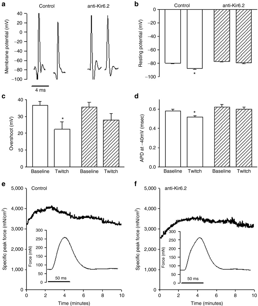
Figure 2.
Effect of Kir6.2 expression inhibition on muscle electrophysiology and force generation in situ. (a) Representative transmembrane action potentials (APs) obtained by floating microelectrode in situ from control (solid bars) versus anti-Kir6.2 (hatched bars) vivo-morpholino treated tibialis anterior (TA) muscles at baseline and following 5–7 minutes of isometric twitching at 1 Hz (twitch). (b) Summary statistics for baseline (control: average for eight mice, 5 APs each and anti-Kir6.2: average for seven mice, 5 APs each) and post-twitch (control: average for seven mice, 10 APs each and anti-Kir6.2: average for seven mice, 10 APs each) resting membrane potential, (c) action potential overshoot, and (d) AP duration at −40 mV (APD−40 mV). Representative tracings of in situ specific peak force over 10 minutes of 1 Hz twitching and single twitch waveforms (insets) from (e) control and (f) anti-Kir6.2 vivo-morpholino treated tibialis anterior. *P < 0.05 for post-twitch versus baseline action potential parameters.

