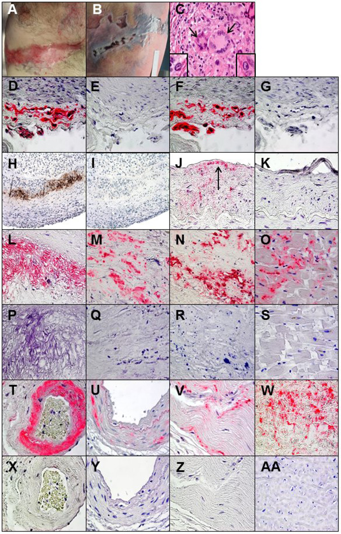
Fig. 1.
Postmortem examination showed linear T6–7 distribution scarred lesions at the site of earlier zoster just below the nipple (A) along with ulcerative necrotic lesions in the same dermatome on the back (B) that were confirmed by PCR to contain VZV. In the right posterior cerebral artery, hematoxylin and eosin staining revealed inflammation and 2 giant cells (C, arrows) in the media adjacent to the disrupted internal elastic lamina, as well as Cowdry A inclusion bodies (C, insets). To confirm the specificity of binding to VZV antigen, immunoperoxidase and immunohistochemical staining were performed with 3 different anti-VZV antibodies. Immunohistochemical staining of a positive control VZV-infected cadaveric cerebral artery with mouse anti-VZV gE IgG1 antibody revealed VZV antigen (D, pink color) that was not seen with mouse isotype IgG1 antibody (E). Immunostaining of the same artery with rabbit anti-VZV IE 63 antibody also revealed VZV antigen (F, pink color) that was not seen with normal rabbit serum (G). In the right posterior cerebral artery, immunoperoxidase staining with a mouse anti-VZV antibody directed against multiple VZV antigens revealed virus in the arterial media (H, brown color) that was not seen when anti-HSV antibody was substituted for anti-VZV antibody (I). Immunohistochemical staining with mouse anti- VZV gE IgG1 antibody also revealed VZV antigen in the thickened arterial intima (J, arrow) that was not seen when adjacent sections were immunostained with mouse isotype IgG1 antibody (K). All other arteries and tissues were immunostained with mouse anti-VZV gE IgG1 antibody, and the presence of VZV antigen in some tissues was confirmed with rabbit anti-VZV IE 63 antibody. Immunostaining with mouse anti-VZV gE IgG1 antibody revealed viral antigen in the media of the aorta (L), the thickened intima of the left anterior descending coronary artery (M), the proximal left circumflex coronary artery (N), the bundle of His (O), the intima and media of an artery adjacent to the adrenal gland (T), the media of the renal artery (U) and the ileum (V), that was not seen when adjacent sections were immunostained with mouse isotype IgG1 antibody (P-S and X-Z). Immunostaining with rabbit anti-VZV IE 63 antibody revealed VZV antigen in sections of the aorta adjacent to that shown in panel L (W), which was not seen when adjacent sections were immunostained with normal rabbit serum (AA). 600× magnification.

