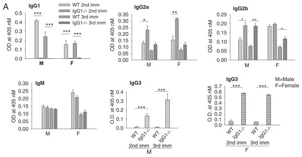
Fig. 2.
Serum AChR antibody levels of IgG1−/− and WT mice. Serum level of anti-AChR auto-Ab was determined by ELISA using affinity purified mouse AChR as a coating antigen. A dramatic increase in anti-AChR IgG3 level was seen in all CFA/AChR immunized IgG1−/− mice (male and female), ***p < 0.001. The increase in serum IgG2a, IgG2b and IgG3, but not IgM anti-AChR Ab was significant in CFA/AChR immunized IgG1−/− mice compared to CFA/AChR immunized wild type mice, *p < 0.05, **p < 0.01. Results are representative of 3 independent experiments. Vertical bars represent standard error, n = 10 each for IgG1−/− and WT mice, M — male; F — female.

