
Figure 1.
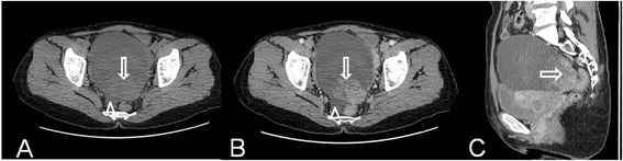
Ovarian clear cell carcinoma in a 49-year-old woman. Axial precontrast MSCT image (A) shows a mostly cystic, unilocular, and oval-shaped mass with smooth margin and irregular solid protrusion (arrow). Axial (B) and sagittal (C) contrast-enhanced CT images show obviously heterogeneous enhancement of the solid component in the tumor (arrow). Note the minimal fluids (arrowhead).