Figure 1.
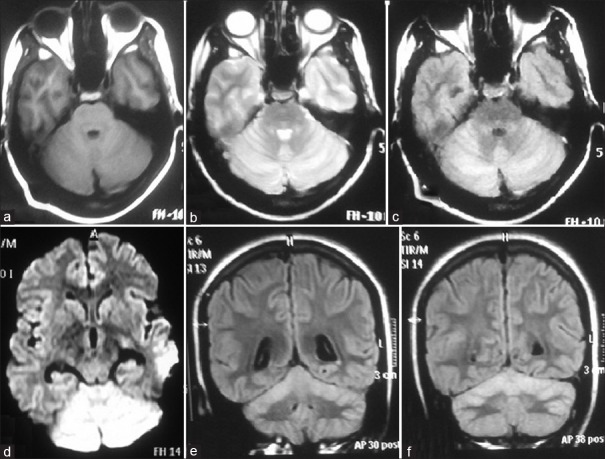
Magnetic resonance imaging (MRI) brain axial images showing bilateral cerebellar hemispheres (a) T1-weighted isointense, (b) T2-weighted hyperintense, (c) fluid-attenuated inversion recovery (FLAIR) hyperintense, (d) restricted diffusion on diffusion-weighted imaging sequence. (e and f) MRI brain coronal FLAIR images showing bilateral cerebellar hyperintense signals

