
Figure 2.
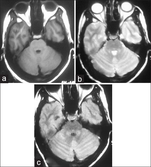
Repeat magnetic resonance imaging brain axial images after 3 months showing partial resolution of the previous hyperintense signals in bilateral cerebellar hemispheres: (a) T1-weighted image, (b) T2-weighted image, (c) fluid-attenuated inversion recovery image