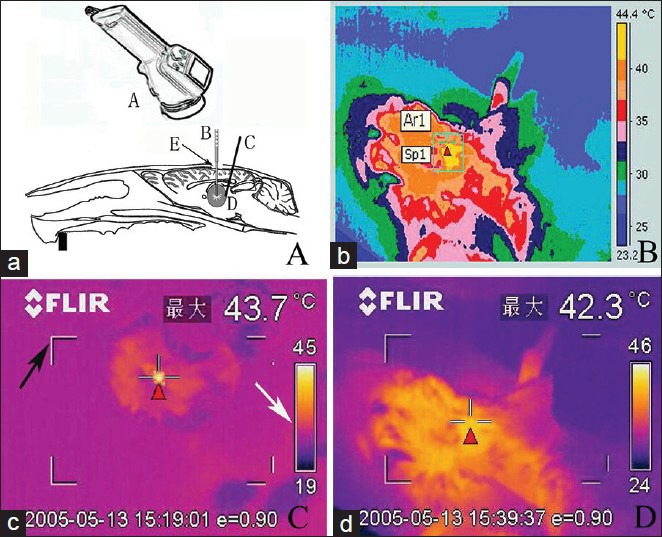
Figure 2.

Experimental rat LITT treatment (a) ThermaCAM S65 type infrared thermometer (A), opened skull window of rat (E). The laser fiber (B) rat brain glioma entity (D), thermocouple electrodes (c) inserted at glioma edge. (b) Image of infrared temperature measurement instrument records. (c) The highest temperature of corresponding regions is displayed, black arrow indicating the viewfinder and white arrow indicating thermal imaging color levels. (d) The target temperature is real-time, as detected on thermal infrared imager, displaying cortex temperature on center target. (Red triangle)
