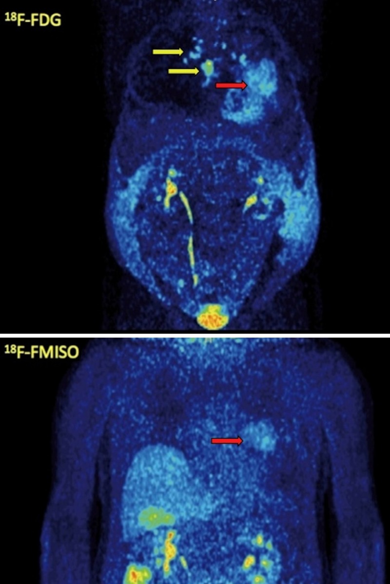
Figure 4.

MIP images of 18F-FDG PET/CT (upper row) and 18F-FMISO PET/CT (lower row) of the same patient as in Figure 3. The patient demonstrates, apart from the primary tumor (red arrow), metastases to contralateral mediastinal lymph nodes and the sternum (yellow arrows), which are depicted, however, only with 18F-FDG PET/CT and not with 18F-FMISO PET/CT. The patient developed also ascites due to the malignancy.
