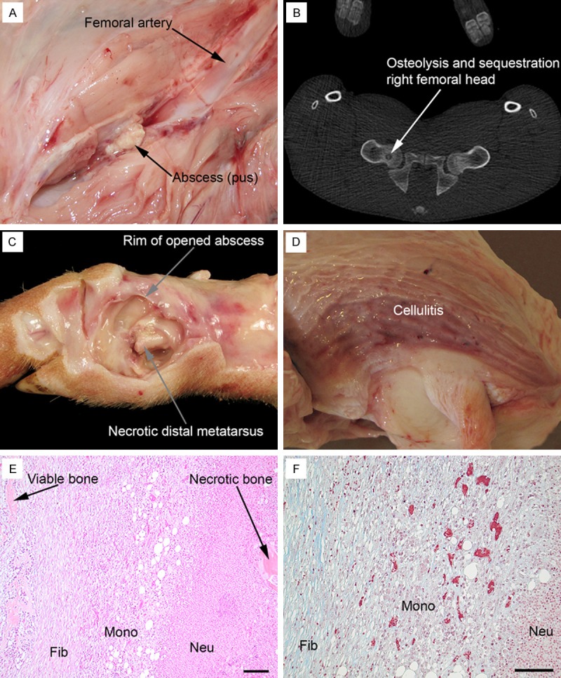
Figure 1.

Gross pathology, CT and histopathology, pig 1, 2 and 4. (A) Small soft tissue abscess, approx. 1 cm in diameter, in the right pelvic limb, pig 1; compare to Figure 3A. (B) CT, transversal projection, of pelvis, pig 1; compare to Figure 3A. (C) Gross pathology of right metatarsus II, medial view, pig 4; the abscess peripheral to the bone has been opened and pus evacuated exposing necrotic metatarsus II bone and a pathological fracture along the growth plate in the distal part of the bone. (D) Cellulitis, i.e. grey-red edematous tissue, peripheral to right patella, pig 2; the cranial surface of patella is present in the lower right part of the picture. Haematoxylin and eosin (E) and Masson trichrome (F) staining (trichrome stains collagen blue) of the tissue bridging viable and necrotic bone components of the right distal metatarsus III, pig 1; fibroblasts (Fib) with collagen formation, mononuclear cells (Mono) and neutrophilic granulocytes (Neu); bar = 100 µm.
