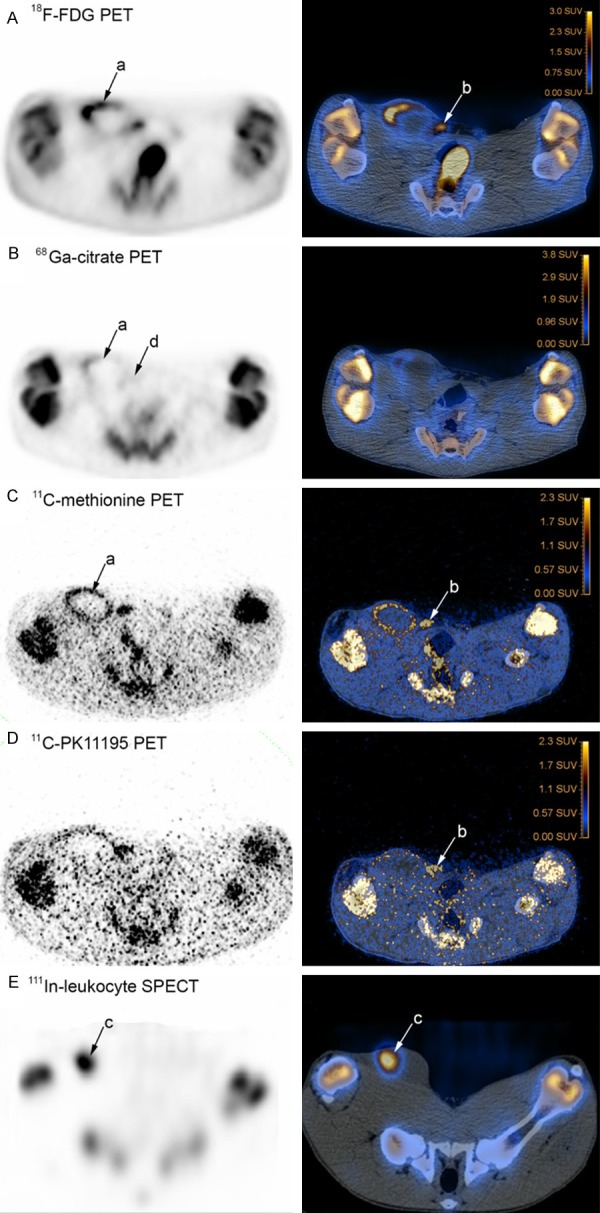
Figure 4.

Scans of pelvic (hind) limbs and region, PET and SPECT to the left, and CT fused images to the right, transversal projections, pig 4. Tracers have accumulated in lesions in the right side of the pig. Sphere-formed accumulations (a) with 18F-FDG (panel A), 68Ga-citrate (panel B) and 11C-methionine (panel C) outline the large, approx. 8 cm in diameter, subcutaneous located abscess; focal accumulation (b) with 18F-FDG (panel A), 11C methionine (panel C) and 11C-PK11195 (panel D) identifies the mammary lymph node, and focal accumulation (c) of 111In-leukocytes (panel E) identify the abscess; with 68Ga-citrate, the mammary lymph node is barely visible (d).
