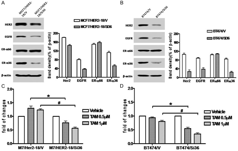
Figure 2.

Knock-down of ER-α36 expression sensitizes HER2 expressing cells to tamoxifen. A. Western blot analysis of the expression levels of ER-α66, ER-α36, HER2 and EGFR in MCF7/HER2-18 cells transfected with an empty expression vector (MCF7/HER2-18/V) and with the ER-α36 shRNA expression vector (MCF7/HER2-18/Si36). B. Western blot analysis of the expression levels of ER-α66, ER-α36, HER2 and EGFR in BT474 cells transfected with an empty expression vector (BT474/V) and with the ER-α36 shRNA expression vector (BT474/Si36). C & D. Cells were treated with indicated concentrations of tamoxifen for seven days and the numbers of survived cells were counted. The columns represent the means of three experiments; bars, SE. * and #, P < 0.05 for control cells transfected with the empty vector vs the cells transfected with ER-α36 the shRNA expression vector, respectively.
