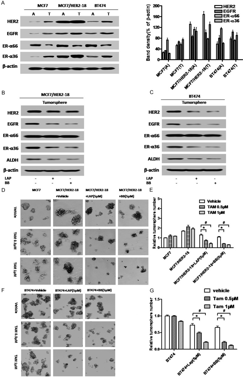
Figure 6.

Disruption of the ER-α36-EGFR/HER2 positive regulatory loops sensitizes HER2 expressing breast cancer stem/progenitor cells to tamoxifen. (A) Western blot analysis of the expression of ER-α36, ER-α66, EGFR and HER2 in the monolayer cells grown on attachment dishes (A) and tumorsphere cells grown on low-attachment dishes (T). Band density (% of β-actin) is also shown. (B & C) Tumorsphere cells derived from BT474 and MCF7/HER2-18 cells were treated with 5 μM of Broussoflavonol B (BB) or Lapatinib (LAP) for five days. Western blot analysis of expression levels different proteins was performed. (D) Tumorsphere formation assay was used to assess the effects of tamoxifen alone or together with Lapatinib (LAP) or Broussoflavonol B (BB) on the breast cancer stem/progenitor cells derived from MCF7/HER2-18 cells. The representative results are shown. (E) The numbers of tumorspheres formed by the cells treated with tamoxifen alone or together with Lapatinib (LAP) or Broussoflavonol B (BB). (F) Tumorsphere formation assay was used to assess the effects of tamoxifen alone or together with Lapatinib (LAP) or Broussoflavonol B (BB) on the breast cancer stem/progenitor cells derived from BT474 cells. The representative results are shown. (G) The numbers of tumorspheres formed by the cells treated with tamoxifen alone or together with Lapatinib (LAP) or Broussoflavonol B (BB). The columns represent the means of three experiments; bars, SE. *&#, P < 0.05 for cells treated with vehicle vs cells treated with 0.5 and 1 μM of tamoxifen.
