Figure 3.

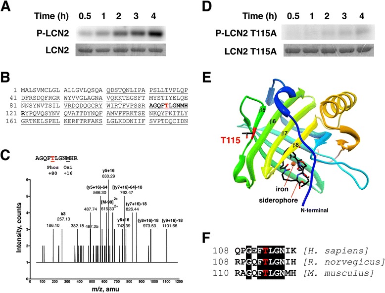
PKCδ phosphorylates LCN2 at T115 in vitro . (A) Recombinant LCN2 proteins were subjected to in vitro kinase assays by PKCδ for up to 4 h. Representative autoradiographs of phosphorylated LCN2 (P-LCN2) are shown in the upper panel. Scanned images of Coomassie blue-stained gels (LCN2) are in the lower panel. (B) Analysis of LCN2 phosphorylation by PKCδ using mass spectrometry. LCN2 was phosphorylated by PKCδ and digested with trypsin. Fragments identified by nano-LC-MS/MS are underlined. The phosphorylated form of peptide AGQF[T]LGNMHR (in bold, amino acids 111–121) was detected. (C) Mass spectrometry spectrum. Identification of the phosphopeptide AGQF[T]LGNMHR (Mr = 1326.57, m/z = 615.33) by tandem MS indicating that the peptide was phosphorylated at T115. (D) Recombinant LCN2 T115A proteins were subjected to in vitro kinase assays by PKCδ for up to 4 h. (E) Crystal structure of LCN2 containing a siderophore (colored by atom type, N = blue, C = black, O = red) and an iron (orange). T115 with side chain shown is located in the β5 strand. (F) Sequence surrounding T115 (highlighted in red) from human, rat, and mouse LCN2 homologs. Conserved amino acids are surrounded by black boxes.
