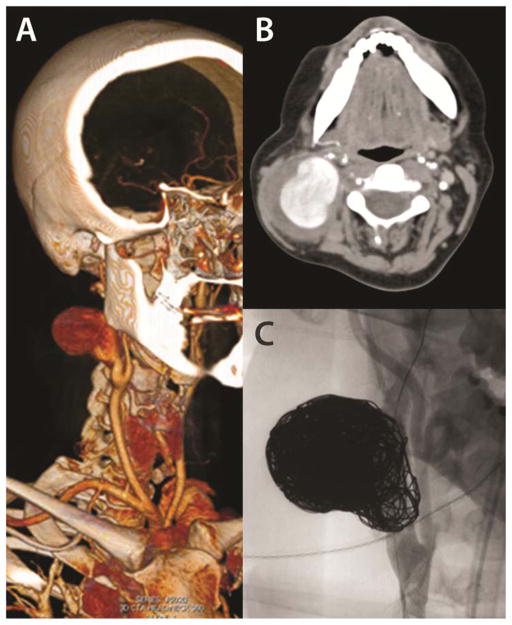
Figure 4.
A 6 cm carotid artery aneurysm which developed in a 34 year old woman acutely in the post-partum period. (A, B) 3-D reconstruction and representative axial image of a computed tomography scan demonstrating the large right internal carotid artery aneurysm. Her arterial pathology led to vEDS diagnosis (MIN mutation, c.1772G>A, p.Gly591Asp) and preoperative multidisciplinary planning accordingly. She was successfully treated with stent-assisted coil embolization of the ICA aneurysm (C). Images courtesy of Johns Hopkins/NIH

