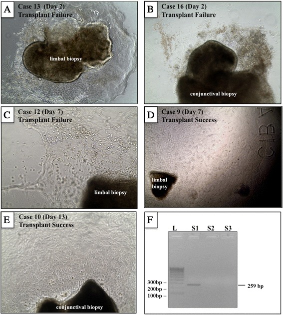
Figure 1.

Cell growth from limbal and conjunctival biopsies. Phase-contrast images of limbal (A, C, D) and conjunctival (B, E) biopsies excised from patients with limbal stem cell deficiency and cultured over a specific period (see panel label for case identification number and time in culture). Although cultures displayed ample proliferation activity, some grafts failed (A-C) whereas others were successful (D and E) at last follow-up. A representative polymerase chain reaction for mycoplasma (F) on conditioned media derived from cultured cells from patient 12 (S2) is shown. S1 (positive control) shows a band at 259 base pairs (bp), and S3 is a negative control.
