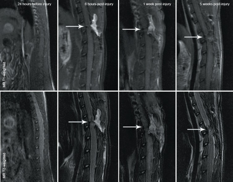
Figure 1.

MRI images of the spinal cord in a rat model of spinal cord transection.
Conventional MR T1- and T2-weighted images show that the signals at the injured spinal cord were decreased after spinal cord transection injury occurred, and the low signals were more common as the time post-injury increased. White arrows refer to the spinal cord injury site.
