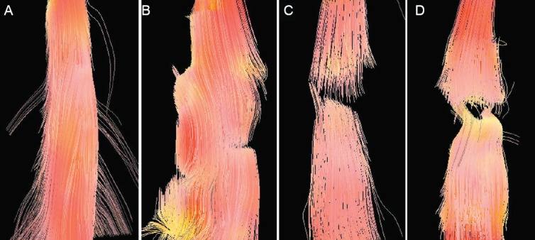
Figure 2.

Fiber tractography images of the spinal cord in a rat model of spinal cord transection.
(A) 24 hours before spinal cord injury, i.e., normal spinal cord; (B) 6 hours post-injury; (C) 1 week post-injury; (D) 5 weeks post-injury. Spinal cord nerve fibers were arranged in a disorderly fashion and the transection gap was widened along with the post-injury time.
