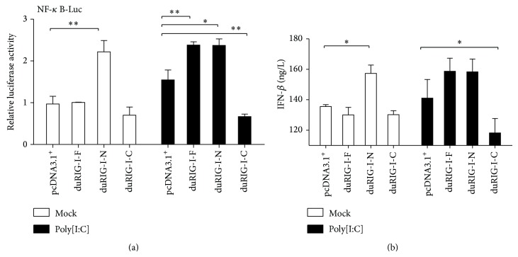
Figure 2.
Characterization of the effect of different domains of duRIG-I on the induction of IFN-β. (a) DF1 cells were transiently transfected with reporter constructs containing pNF-κB-Luc and internal control vector pRL-TK together with empty vector pcDNA3.1+, duRIG-I-F, duRIG-I-N, and duRIG-I-C. The effector/reporter/internal control ratio was 4 : 1 : 1/15. Transfected cells were mock treated (Mock), treated with poly[I:C] for 12 h, were analyzed by the dual-luciferase assay. The data represent relative luciferase activity, normalized to Renilla luciferase activity. The error bars represent SE of triplicate transfections. (b) IFN-β in the culture medium was quantified by ELISA after collection of the culture supernatant.

