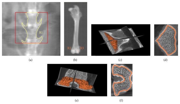
Figure 1.
DXA and μCT measurement of vertebrae and femoral condyles. (a) DXA measurement of vertebrae. R1: ROI of L3 and L4. (b) DXA measurement of femoral condyles. R2 and R3: ROI of femoral condyles (7 mm × 7 mm ROI). (c) The 3D reconstruction of the ROI in vertebrae in μCT measurement. (d) The ROI on the axial section of vertebrae in μCT measurement. (e) The 3D reconstruction of the ROI in femoral condyles in μCT measurement. (f) The ROI on the axial section of femoral condyles in μCT measurement. DXA: dual-energy X-ray absorptiometry; ROI: regions of interest.

