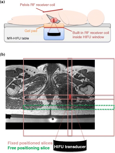
Figure 1.

Slice positioning of the MRT scans. An example of an MR-HIFU setup for a treatment in the pelvis is shown in (a). An example of the MRT scan slice positioning is shown in (b), on a T1-weighted planning scan of an osteolytic lesion in the pubic bone (treatment 10). Three slices (light-red) were fixed with the centers to the location of the HIFU focus; one slice could be freely placed by the user and was placed in the near-field area of the HIFU beams (green, dashed).
