
Figure 1.
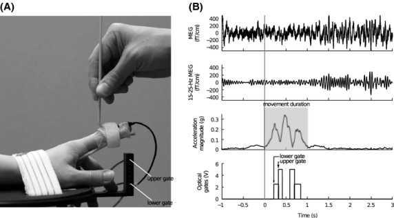
(A) The arrangement for passive movement. (B) Representative signals of one subject during right index finger passive movement. Two upper rows: MEG signal from a single gradiometer channel (raw and filtered 15–25 Hz) over the primary sensorimotor cortex. The ∽20-Hz modulation of the filtered MEG signal is observable even to a single movement. Third row: magnitude of acceleration (i.e., the Euclidean norm of the three accelerations). Total duration of movement is highlighted with gray. Lowest row: trigger signals from the lower (1st) and upper (2nd) optical gates.