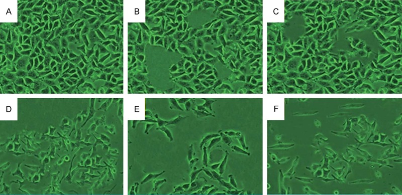
Figure 5.

Cell apoptosis observed by Hoechst 33342 staining. HeLa cells were treated with (B) 60 μM myricetin (MYR), (C) 60 μM methyl eugenol (MEG), (D) 1 μM cisplatin (CP), (E) combination of MYR + CP and (F) combination of MEG + CP for 48 h. (A) Compared with the control group, treated cells exhibited chromatin condensation, nuclear fragmentation and apoptotic body formation.
