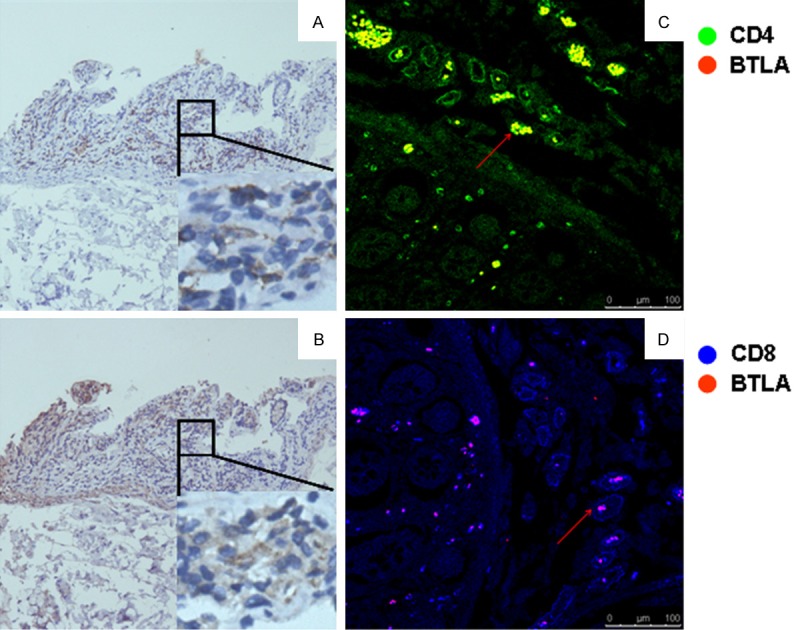
Figure 1.

BTLA expression in colon tissue from the patient with UC. Colon tissues from UC patients were stained with enzyme- or fluorescence-labeled antibodies, and then observed under a light microscopy or Laser Scanning Confocal Microscopy. Same area in colon tissue were stained with anti-human CD3 (A) or anti-human BTLA (B) respectively, immunohistochemically. (C) and (D) show co-staining of BTLA (red) with CD4 (green) or CD8 (blue).
