
Figure 5.
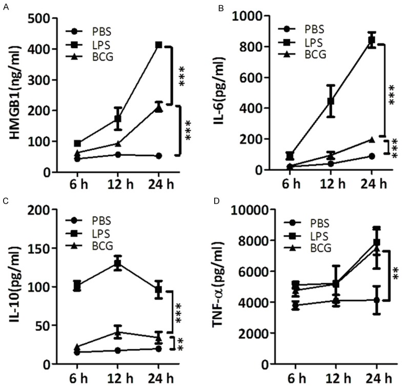
Abundance of HMGB1, IL-6, IL-10 and TNF-α in supernatant of THP-1 cells after BCG infection. PMA differentiated THP-1 cells were infected with BCG. Infection was continued for 6, 12 and 24 hrs and supernatant was collected for the detection of HMGB1, IL-6, IL-10 and TNF-α. LPS was used as a positive control and PBS was used as a negative control. The values and error bars represent average and standard error of the mean (SEM) of three independent set of experiments. *, P<0.05; **, P<0.01; ***, P<0.0001.