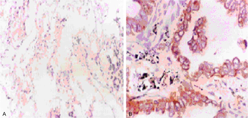
Figure 1.

Immunohistochemistry analysis of BI-1 expression in clinical lung samples. All positively stained cells were of epithelial cells origin and showed a brown cytoplasmic staining around the clearly demarcated nuclei. Very weak, diffuse staining of BI-1 was detected in normal tissue (A), while strong staining of BI-1 was detected in tumor tissue (B).
