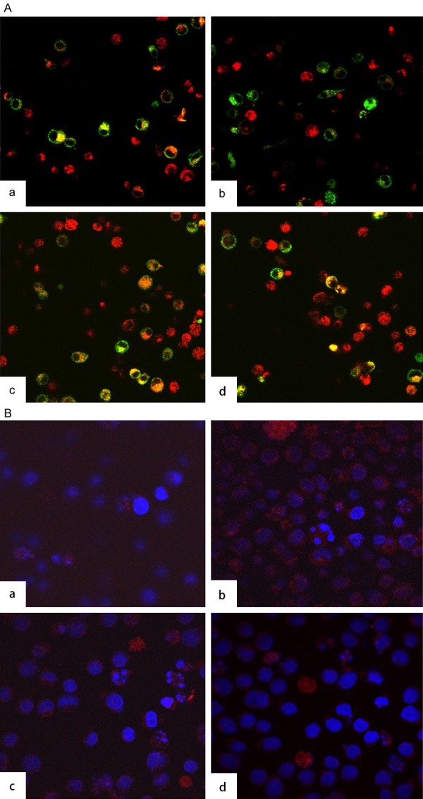
Figure 6.

Co-localization and In situ PLA reveals interaction of CD63 and HIV-1 Gag. A. Co-localization of CD63 and HIV-1 Gag in U1 cells. U1/HIV-1 cells (2 × 10⁶ cells/well) were plated in triplicate at 6-well plates and transfected with 50 nM siRNAs (CD63, ERBB2IP and TSG 101). Controls included cells induced. Forty eight hours post-transfection, cells were treated with 5 × 10⁻⁶ M PMA. Cells were fixed on day 2 post PMA induction, followed by double staining of CD63 and HIV-1 Gag with PE-labelled anti-CD63 and FITC labelled anti-HIV Gag. Confocal images showed co-localization of CD63 with HIV-1 Gag in (a) U1 cells, (b) CD63 siRNA transfected cells, (c) ERBB2IP siRNA transfected cells, and (d) TSG101 siRNA transfected cells, respectively. B. Detection of CD63 and Gag 17 in U1 cells using Duolink with two primary antibodies. U1/HIV-1 cells were treated with (a) CD63 specific siRNA; (b). ERBB2IP siRNA; (c) Tsg 101 siRNA; (d) No treatment, two primary antibodies were anti-CD63 and anti-integrin α4 as positive control. Two primary antibodies for a, b, and c were anti-CD63 and anti Gag 17.
