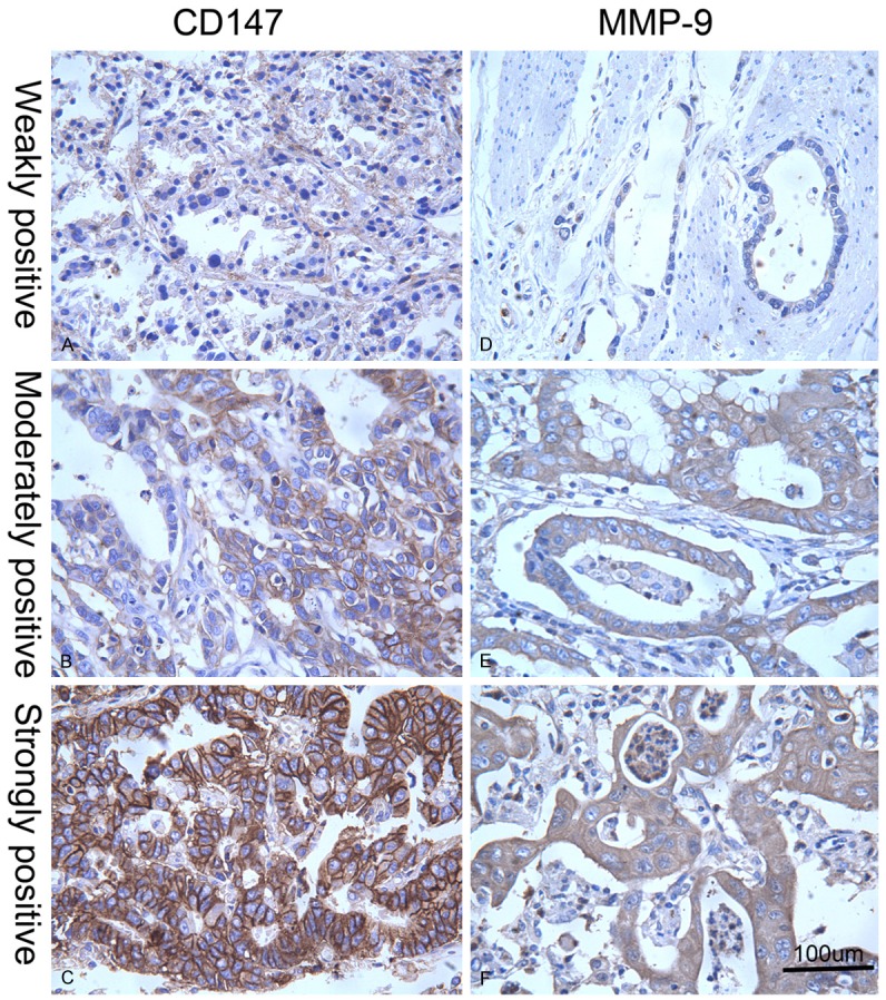
Figure 2.

Different expression levels of CD147 and MMP-9 in AEG tissues. Revealed by immunohistochemical staining in AEG tissues, weakly positive (A), moderately positive (B), and strongly positive (C) CD147 expressions were indicated by yellow or brown color in cytoplasm or on cellular membrane, and weakly positive (D), moderately positive (E), and strongly positive (F) MMP-9 expressions were illustrated by brown color in cytoplasm (high power field, × 400). MMP-9, matrix metalloproteinase 9; AEG, adenocarcinoma of esophagogastric junction.
