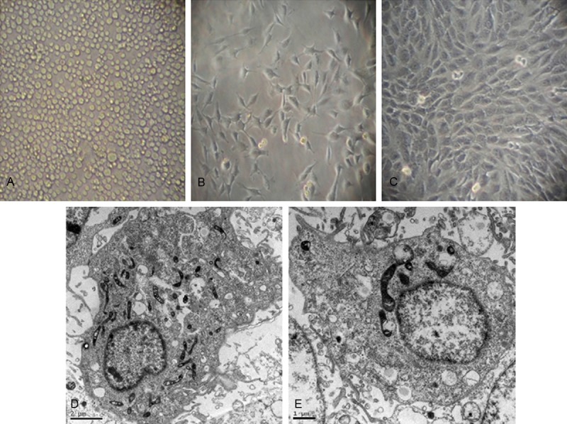
Figure 2.

Morphological characteristics of endothelial progenitor cells derived from bone marrow mononuclear cells. Light microscopy of bone marrow mononuclear cells isolatd from SD rats. (A) Freshly isolated bone marrow mononuclear cells. (B) Early outgrowth of endothelial progenitor cells by day 7 of primary culture. The cells are fusiform, triangular, or irregular in shape. (C) Endothelial progenitor cells exhibit a typical cobblestone-like appearance by day 21. Transmission electron microscopy shows abundant finger-like, spherical, or villous protrusions on the surface of the endothelial progenitor cells (D) and Weible-Palade (W-P) corpuscle (E).
