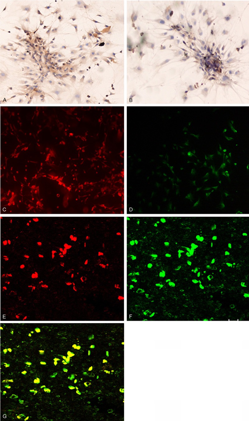
Figure 3.

Immunohistochemical staining of cultured bone marrow mononuclear cells at day 7 for vWF (A) and VEGFR-2 (B). Immunofluorescence staining for CD34 (C) and CD133 (D). Magnification × 200 Confocal microscopy showed (E) EPCs after the cytoplasm of EPCs took up the DiL-Ac-LDL stain (shown in red), (F) EPCs stained positive for FITC-UEA-1 (shown in green), and (G) EPCs with double positive staining for phagocytic DiL-Ac-LDL and FITC-Lectin-UEA-1 (shown in yellow). Magnification: × 200.
