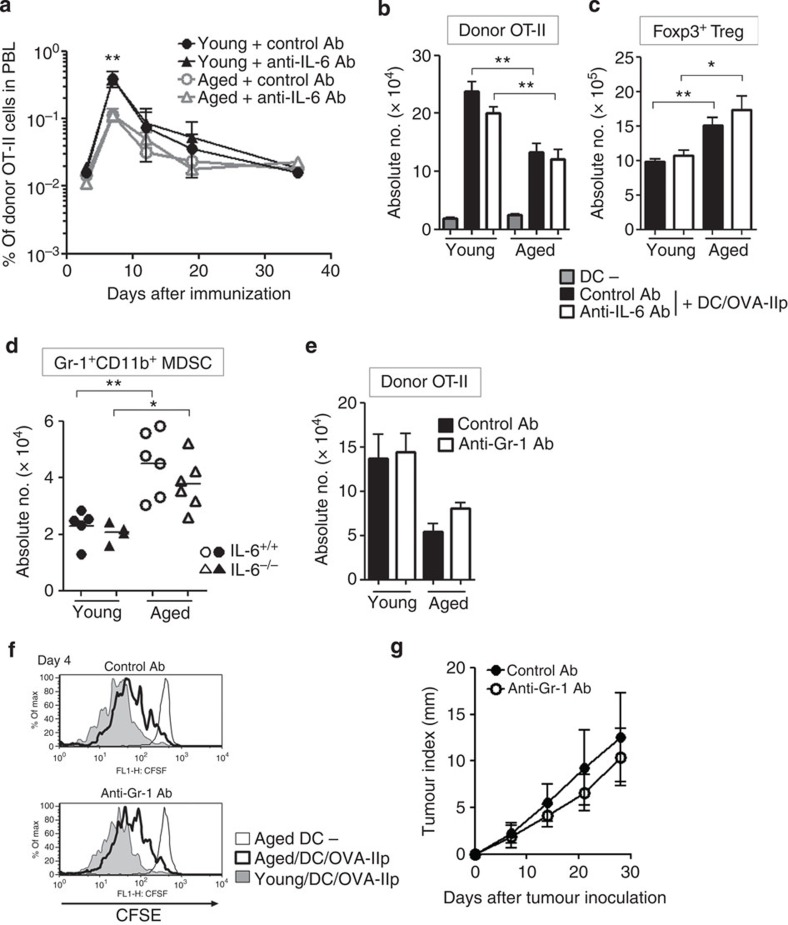
Figure 3. The aged environment dampens antigen-induced proliferation of CD4+ T cells.
(a–c) OT-II cell transfer, immunization and Ab treatment were performed as described in Fig. 1d. The frequency of donor CD45.1+OT-II cells in PBL was monitored over time (a), and the total number of donor OT-II cells (b) and CD4+Foxp3+Treg cells (c) in spleen and LNs were determined 6 days after immunization. (d) The number of Gr-1+CD11b+ MDSC in spleen and LN cells from young or aged IL-6+/+ or IL-6−/− mice were determined. (e–g) Anti-Gr-1 Ab was injected 5 days before transfer of the CFSE-labelled OT-II cells, followed by immunization. The total number at day 6 (e) and CFSE profile at day 4 (f) of donor OT-II cells under the indicated conditions are shown. Seven days after immunization, MCA-OVA were inoculated into the aged mice. The outgrowth in aged mice was measured over time (g). The values are mean±s.e.m. (n=6 per group); *P<0.05, **P<0.01, analysis of variance followed by Tukey's post hoc test. Representative data from two or three experiments with similar results are presented.

