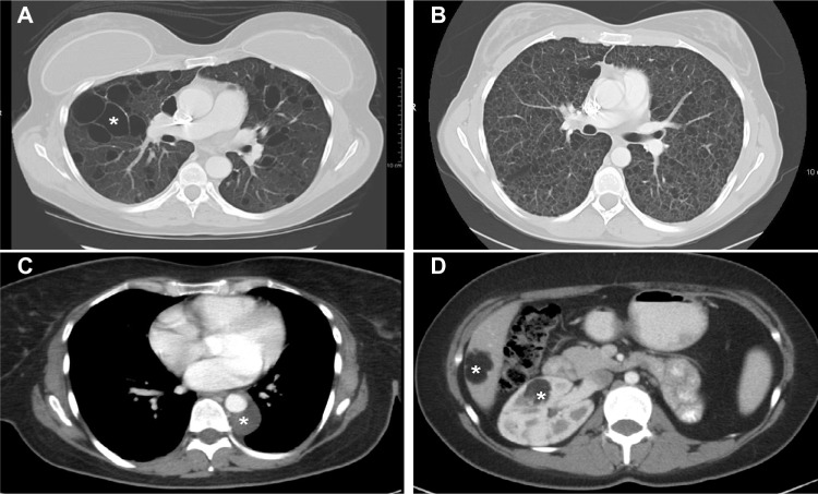
Figure 3.
Computed tomography scans showing pulmonary and extrapulmonary images of patients with LAM.
Notes: (A) Multiple large (white asterisk) thin-walled cysts scattered throughout the lungs. Between the cystic areas there is normal-appearing lung parenchyma. (B) Numerous small thin-walled cysts have completely replaced the normal lung parenchyma. (C) Small posterior mediastinal lymphangioleiomyoma surrounding the descending aorta (white asterisk). (D) Right renal (white asterisk) and liver (white asterisk) angiomyolipomas in a patient with TSC-LAM. The fatty, low-density component is clearly visualized.
Abbreviations: LAM, lymphangioleiomyomatosis; TSC, tuberous sclerosis complex.

