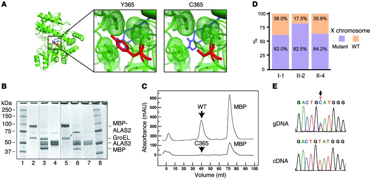
Figure 2. Severe LOF with the ALAS2 Y365C mutation and lack of highly skewed X inactivation in female mutation carriers.
(A) Model of ALAS2 shows PLP highlighted in blue and the Y or C amino acid at position 365 highlighted in red. (B) SDS-PAGE gel of WT and mutant ALAS2. Lanes 1 and 8 contain the protein standards, while lanes 2–4 and 5–7 contain WT and mutant ALAS2 protein samples, respectively. Lanes 2 and 5 show partially purified samples after amylose affinity chromatography, lanes 3 and 6 show results after factor Xa digestion, and lanes 4 and 7 show results after gel filtration chromatography. Thin vertical lines in this composite figure separate noncontiguous lanes in the 2 original gels. (C) Chromatographic profiles for purification of WT and mutant ALAS2 by size exclusion (absorbance at 280 nm is shown in milliabsorbance units [mAU]). (D) Quantification of HUMARA results in all affected individuals showing WT and mutant X chromosomes. (E) Sanger sequencing traces of genomic DNA (gDNA) and cDNA derived from reticulocyte mRNA from the proband (II-2) for ALAS2, with an arrow highlighting the mutation.

