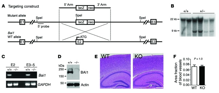
Figure 1. Generation and characterization of Bai1–/– mice.
(A) Schematic of the Bai1 gene–targeting strategy. Homologous recombination of the targeting construct with the mouse Bai1 locus results in deletion of exon 2 (E2), which leads to a null mutation. (B) Southern blot genotyping of targeted ES cell clones. Genomic DNA from G418-resistant clones was digested with SpeI. After electrophoresis, the digested DNA was transferred to membrane and hybridized with the 5′ probe. The WT allele migrated as a 22-kb fragment, while the correct homologous recombinant allele migrated as a 9-kb fragment. (C) RT-PCR for Bai1 gene transcripts from either exon 2 (E2) or exons 3–5 (E3–5) in 2-week-old WT and KO brains. (D) WB analysis to confirm the absence of BAI1 protein in homozygous KO (–/–) mouse brains. (E) H&E staining of brain sections showed no apparent structural defects. Scale bar: 0.5 mm. (F) Quantification of vasculature in the brain showed no difference between WT and KO mice (n = 6 mice/group; P = 1.0 by 2-tailed Student’s t test).

