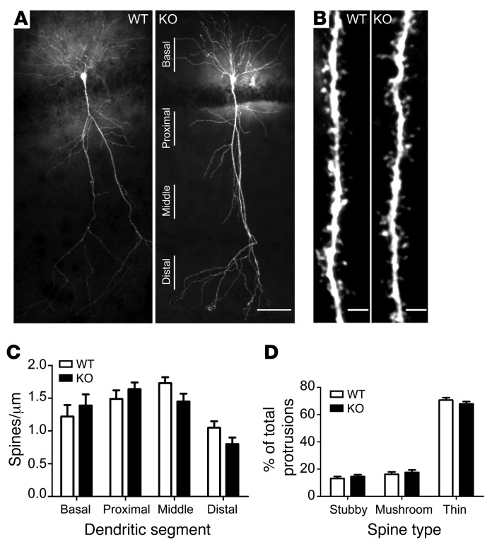
Figure 4. Bai1–/– hippocampal CA1 neurons have normal dendritic arborization and spine morphology.
(A) Representative images of biocytin-filled CA1 neurons from adult WT and KO mice show normal dendritic arborization in KO mice. Scale bar: 50 μm. (B) Representative images from apical segments of WT and KO neurons show unaffected dendritic spine density and morphology. Scale bars: 2 μm. (C) No significant difference in dendritic spine density was observed between WT and KO CA1 neurons. Secondary dendritic segments were captured from basal dendrites, proximal apical dendrites (50–100 μm from the soma), middle apical dendrites (100–150 μm from the soma), and distal apical dendrites (>150 μm from the soma). n = 5 mice/group; P = 0.55. (D) Dendritic spine morphology was not affected in KO neurons. Spines were classified as stubby, mushroom, or thin and were quantified. n = 5 mice/group; P = 0.99. All data represent the mean ± SEM and were analyzed by 2-way ANOVA.

