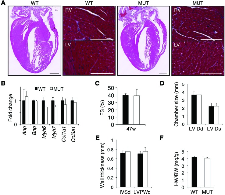
Figure 2. No overt changes in cardiac remodeling, function, hypertrophy, or the fetal gene program were observed in the FuseNax mice.
(A) Four-chamber views of Masson’s Trichrome–stained hearts taken from 4-month-old WT and FuseNax (MUT) mice. Scale bar: 1 mm. Higher-magnification images of right ventricles (RV) and left ventricles (LV) are shown to the right of respective 4-chamber views. Note no significant morphological changes or increase in fibrosis. n = 3 mice per genotype. Scale bar: 100 μm. (B) qRT-PCR analysis of the indicated genes showing the fold change relative to 18S from 10-month-old mice. Note that expression of the fetal gene program (Anp, Bnp, αMHC, βMHC) and fibrotic markers (collagen 1A1, collagen 3A1) were unchanged in FuseNax mice compared with those in WT mice. Data represent mean ± SEM; n = 8 mice per genotype. (C–E) Fractional shortening (FS) was calculated, and chamber size and wall thicknesses were measured in 47-week-old mice using echocardiography. Note that no changes were observed in heart function. Data represent mean ± SD, n = 16 mice per genotype. LVIDd, left ventricle internal diameter, diastole; LVIDs, left ventricular internal diameter, systole; IVSd, interventricular septum diameter; LVPWd, LV posterior wall, diastole. (F) Heart weight/body weight ratios were unaffected in the FuseNax mice. Data represent mean ± SEM; n = 16 mice per genotype. Two-tailed Student’s t test was performed on data.

