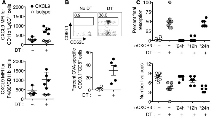
Figure 10. CXCR3 blockade protects against fetal wastage and decidual fetal-specific CD8+ T cell accumulation triggered by partial depletion of maternal FOXP3+ Tregs.
(A) Mean fluorescent intensity after staining with anti-CXCL9 compared with isotype control antibody among neutrophils (CD11b+ Ly6Cint) and macrophages (F4/80+ CD11b–) recovered from the decidua 3 days after initiating DT treatment to E11.5 Foxp3DTR/WT female mice on the C57BL/6 background bearing allogeneic pregnancies after mating with BALB/c-OVA males. (B) Representative FACS plots and composite data showing the percentage of fetal-OVA257–264-specific cells (CD90.1+) among CD8+ T cells recovered from the decidua 3 days after initiating DT treatment compared with mice with no DT treatment described in A. (C) Percentage of resorbed fetuses and number of live pups for Foxp3DTR/WT female mice on the C57BL/6 background bearing allogeneic pregnancies after mating with BALB/c males that were administered anti-CXCR3 antibody (500 μg per mouse) 24 hours before or 12 or 24 hours after initiating sustained daily DT treatment (E11.5), compared with controls with no DT or anti-CXCR3 antibody treatment. Each symbol indicates the data from a single mouse, and these results, containing 3 to 8 mice per group, are representative of 3 independent experiments, each with similar results. Error bars represent mean ± 1 SEM.

