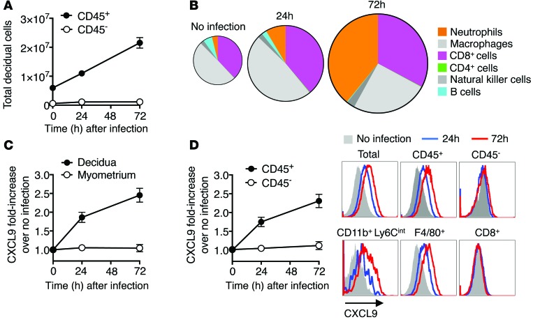
Figure 4. CXCL9-producing inflammatory cells accumulate in the decidua after prenatal L. monocytogenes infection.
(A) Number of CD45+ leukocyte and CD45– nonleukocyte stromal cells recovered from the decidua at each time point after L. monocytogenes ΔactA (107 CFU) infection initiated midgestation (E11.5) among C57BL/6 female mice during allogeneic pregnancies after mating with BALB/c males. (B) Pie chart illustrating quantitative accumulation and quantitative shifts in each CD45+ leukocyte subset recovered from the decidua for mice described in A. Individual leukocyte subsets were delineated after gating on CD45+ cells and identified as neutrophils (CD11b+Ly6Cint), macrophages (F4/80+CD11b–), natural killer cells (NK1.1+CD4–CD8–), B cells (B220+CD4–CD8–), CD4 cells (CD4+CD8–), and CD8 cells (CD8+CD4–). (C) Relative CXCL9 expression among cells recovered from the decidua compared with adjacent myometrium after L. monocytogenes ΔactA (107 CFU) infection for mice described in A. (D) Relative CXCL9 expression among CD45+ compared with CD45– decidual cells and representative histogram plots showing CXCL9 expression by each cell type before (gray shaded) and 24 (blue line) or 72 (red line) hours after L. monocytogenes ΔactA infection. These data showing average results from 5 to 10 mice per group per time point are representative of 3 independent experiments, each with similar results. Error bars represent mean ± 1 SEM.

