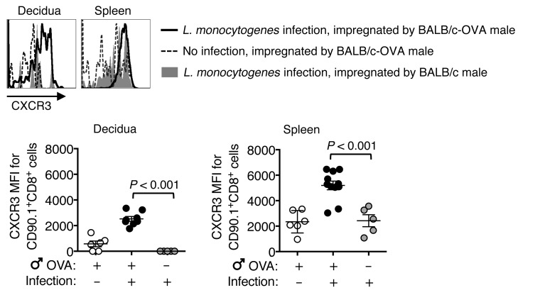
Figure 6. Prenatal L. monocytogenes infection selectively primes CXCR3 expression by maternal CD8+ T cells with fetal specificity.
Representative plots and composite analysis showing relative expression of CXCR3 by OVA257–264-specific (CD90.1+) CD8+ T cells recovered from the decidua or spleen among C57BL/6 female mice bearing allogeneic pregnancies after mating with BALB/c-OVA mice compared with BALB/c males 3 days after L. monocytogenes ΔactA (107 CFU) infection initiated midgestation (E11.5) and controls without infection. Each symbol reflects the data from a single mouse, and these results, containing 5–11 mice per group, are representative of 3 independent experiments, each with similar results. Error bars represent mean ± 1 SEM. Differences between the indicated groups were evaluated using the 1-way ANOVA statistical test. MFI, mean fluorescent intensity.

