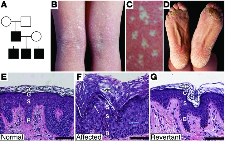
Figure 1. IWC pedigree, clinical features, and histology of affected and revertant skin.
(A) Pedigree. (B) Index case popliteal fossa shows numerous revertant spots. (C) Revertant clones have a phylloid configuration, with intervening focal affected skin islands. (D) Thick scale on the feet. Histology of (E) normal, (F) affected, and (G) revertant skin, showing basal layer (B), spinous layer (S), granular layer (G), and stratum corneum (SC). Affected skin shows hypercellularity, increased epidermal thickness, prominent keratohyalin granules (white arrow), and perinuclear vacuolization with rare binucleate cells (blue arrow). Revertant skin shows normal histology. Scale bar: 50 mm.

