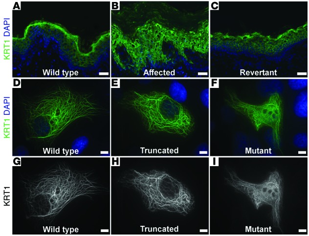
Figure 3. Mutant KRT1 filaments are mislocalized in vivo and in vitro.
(A–C) Immunolocalization of KRT1 reveals collapsed filament networks in affected skin (B) visible as bright perinuclear rings, which are absent in (A) normal and (C) revertant skin. (D–I) Immunolocalization of KRT1 in primary liver hepatoma cells transfected with (D) wild-type KRT1, (E) KRT1 truncated at the site of the frameshift identified in the index case, and (F) mutant KRT1IWC. Wild-type and truncated KRT1 integrate into the cytoplasmic filament network, while cells expressing KRT1IWC show filament network collapse and nuclear accumulation. (G–I) KRT1 staining alone reveals that (I) the mutant protein accumulates in the nucleus. Scale bars: 50 mm (A–C); 5 mm (D–I).

