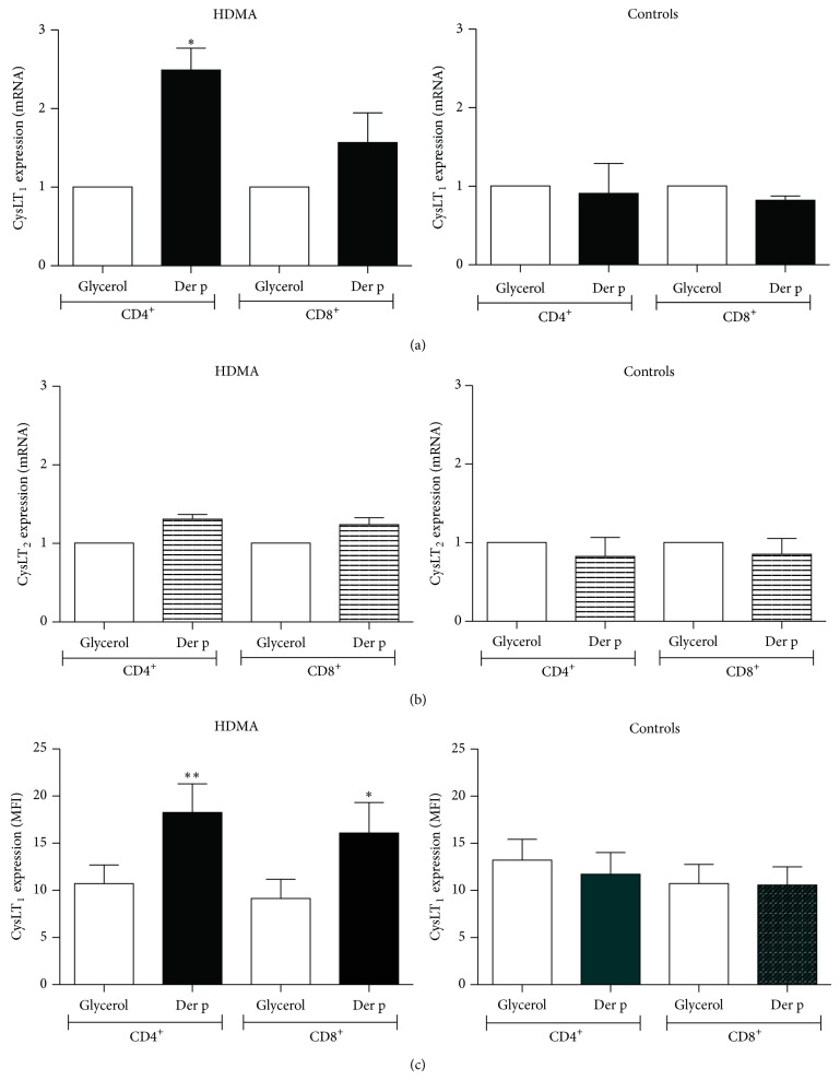
Figure 1.
Der p effect on T cell CysLT1 and CysLT2 mRNA and protein expression. Comparison of CysLT1 and CysLT2 mRNA expression in CD4+ and CD8+ T cells from healthy controls and HDM-allergic (HDMA) patients following stimulation with the Der p allergen (200 AU/mL). PBMC from healthy control and HDMA subjects were cultured for 48 h (qPCR) or 72 h (FACS) in the presence of glycerol vehicle or Der p before CD4+ and CD8+ T cells were purified and collected for analysis. CysLT1 (a) and CysLT2 (b) mRNA expression was measured by real-time quantitative PCR analysis. Data are presented as fold (ΔΔCt) increases over GAPDH mRNA (±SEM). ∗ P < 0.05 and ∗∗ P < 0.01, relative to vehicle glycerol; n = 6 for controls; n = 10 for HDMA. Cell surface expression of CysLT1 (c) receptor was evaluated using rabbit polyclonal anti-CysLT1 receptor Ab, followed by labeling with FITC-conjugated goat anti-rabbit IgG. Cells were further incubated with anti-CD4 PE-Cy5 and anti-CD8 PE Ab before analysis on a FACSCalibur flow cytometer. Data are expressed as geometric mean (±SEM) fluorescence intensity (MFI). ∗ P < 0.05; n = 6 for controls; n = 10 for HDMA.

