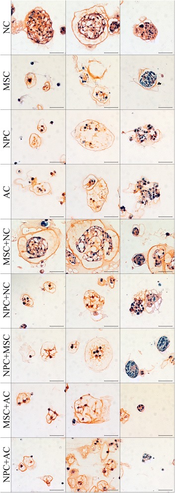
Figure 2.

Extracellular matrix deposition. Histopathological slides of typical cell morphologies on day 28 of notochordal cells (NCs), mesenchymal stromal cells (MSCs), nucleus pulposus cells (NPCs), articular chondrocytes (ACs), MSC + NC, NPC + NC, NPC + MSC, MSC + AC, and NPC + AC. Prior to staining, alginate was removed with sodium citrate. Cell nuclei are stained blue (hematoxylin), proteoglycans are red (Safranin O) and collagen is green (Fast Green) (scale bar = 50 μm).
