Figure 5.
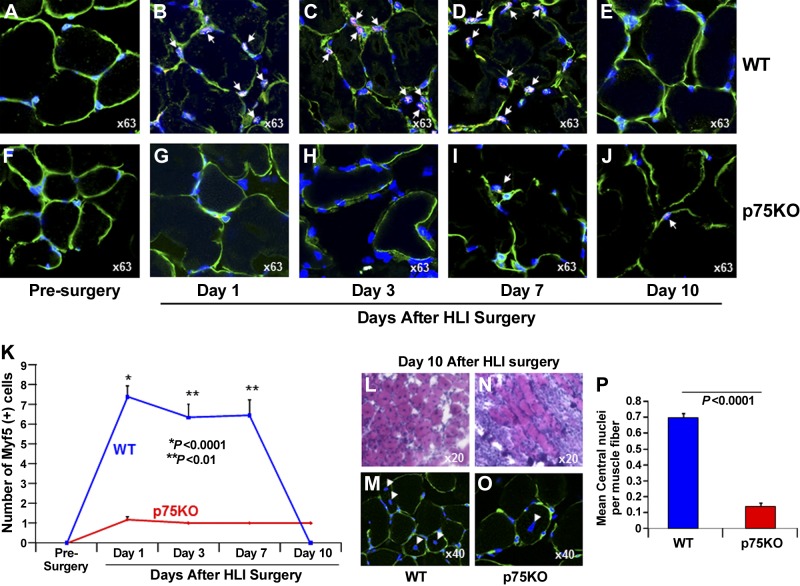
Ischemia-induced satellite-cell activation is impaired in young p75KO mice. Representative ×63 confocal microscopy images for triple immunostaining with satellite-cell marker: Myf5 (red), laminin (green) to visualize the basement membrane, and TO-PRO-3 (blue) to identify nuclei in HL muscle tissue from WT and p75KO mice, respectively, at (A, F) presurgery control, (B, G) d 1 after HLI surgery, (C, H) d 3 after HLI surgery, (D, I) d 7 after HLI surgery, and (E, J) d 10 after HLI surgery. White arrows indicate Myf5/TO-PRO-3+ satellite cells in the muscle tissue, and the green laminin staining identifies the basement membrane. K) Graphic representation of number of Myf5+ cells in the HL muscle tissue isolated from WT and p75KO mice before surgery and up to 10 d after HLI surgery. Representative ×20 bright field microscopy images of H&E staining in HL muscle tissue from (L) WT and (N) p75KO mice 10 d after HLI surgery. Representative ×40 confocal images of laminin (green) and TO-PRO-3 (blue) double staining in HL muscle tissue from (M) WT and (O) p75KO to confirm migratory properties and centralized of myonuclei in WT and p75KO muscle fibers on d 10 after HLI surgery. N) Graphic representation of mean number of central nuclei/muscle fiber in the HL muscle tissue isolated from WT and p75KO mice. All results are presented as means ± sem; n = 3–5 animals per time point per group for WT (blue bar) and p75KO (red bar) groups. Statistical significance was assigned when P < 0.05.

