Figure 1.
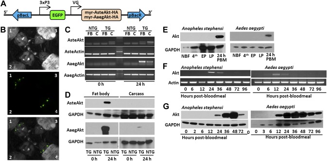
Generation and expression profiles of TG A. stephensi and Ae. aegypti mosquitoes with increased IIS in fat body. A) Schematic of construct engineered into TG mosquito lines. Myristoylated Akts were linked to an HA epitope to facilitate protein identification and engineered downstream of the A. gambiae or Ae. aegypti VG (VG) promoter. The EGFP fluorescent marker was linked to the synthetic 3XP3 promoter to drive expression in the eyes and nervous system. These 2 genes were flanked by the left and right arms of the piggyback (pBac) transposon. B) Example of EGFP expression in the eyes of larval TG Ae. aegypti (1) and A. stephensi (4) compared to NTG Ae. aegypti (2) and A. stephensi (3). The top panel shows the larvae under white light, the middle with a green fluorescent protein filter, and the third a merging of the top 2. C) Transcript expression in the fat body (FB) and carcass (C) of TG and NTG mosquitoes before blood feeding (0 hours) and 24 hours after blood feeding (24 hours). Actin was used as a positive control. D) Protein expression in the fat body and carcass of TG and NTG mosquitoes before blood feeding (0 hours) and 24 hours after blood feeding (24 hours). E) Expression profile of myr-AsteAkt-HA protein during mosquito development. Non-blood-fed adult females (NBF), fourth-instar larva (4th), early pupae (EP), late pupae (LP), and 24 hours after blood meal for A. stephensi (AsteAkt) and Ae. aegypti (AaegAkt). F) Postblood meal time course of transcript expression in the fat body of adult TG A. stephensi females utilizing myr-AsteAkt-specific primers (top) showed that transcript expression occurred from 12 to 36 hours after blood meal. AsteActin-specific primers were used as a positive control. Postblood meal time course of protein expression in the fat bodies of TG A. stephensi females (bottom) shows that myr-AsteAkt-HA protein is first present 24 hours after blood meal, reaches maximal expression between 36 and 48 hours after blood meal, and begins to decline by 72 hours after blood meal. GAPDH protein expression was used as a loading control. G) Postblood meal time course of transcript expression in the fat body of TG Ae. aegypti mosquitoes utilizing myr-AaegAkt-specific primers (top) showed that expression occurred at 12 and 24 hours after blood meal. AaegActin-specific primers were used as positive control. Postblood meal time course of protein expression in the fat bodies of TG A. stephensi females (bottom) shows that myr-AsteAkt-HA protein is first present 12 hours after blood meal, reaches maximal expression between 24 and 48 hours after blood meal, and begins to decline by 96 hours after blood meal. GAPDH was used as a loading control for protein analysis. All transcript and protein expression studies were replicated a minimum of 3 times.

