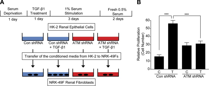
Figure 8.
Increased renal fibroblast proliferation by conditioned media derived from TGF-β1–stimulated con shRNA-, but not ATM shRNA-, expressing HK-2 cells. A) Schematic of epithelial-fibroblast cross-talk. Con shRNA or ATM-depleted HK-2 cells were treated with TGF-β1 for 1 d followed by additon of 1% serum for 3 d to promote growth arrest, washed with PBS to remove any residual TGF-β1 prior to addition of fresh 0.5% FBS/DMEM for 48 h. Conditioned media isolated from epithelial cells were directly added to semiconfluent NRK-49F fibroblasts at similar cell density for 2 d. B) Plots (mean ± sd) represent relative NRK-49F cell counts of over 100 (2 × 2 mm) fields for each experimental condition. ***P < 0.001.

