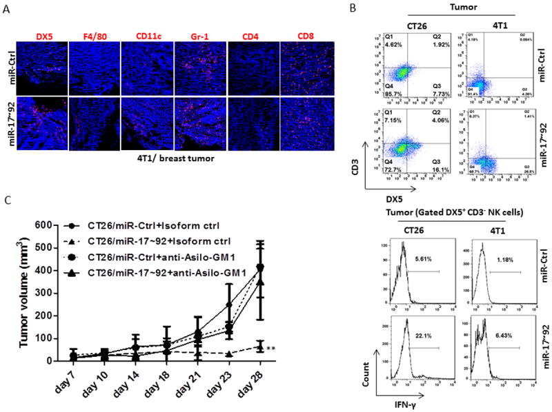
Figure 2. NK cells play critical roles in the suppression of murine breast and colon tumor growth in vivo.

(A) Immunofluorescent staining showing DX5+, F4/80+, CD11c+, Gr-1+, CD4+, and CD8+ immune cells infiltrated into tumors at 28 days after orthotopic injection of 4T1/miR-Ctrl or 4T1/miR-17~92 monoclonal cells into the mammary fat pads in BALB/c mice.
(B) Leukocytes were isolated from 28-day tumor after injected orthotopically with 4T1/miR-Ctrl and 4T1/miR-17~92 monoclonal cells into the mammary fat pads or subcutaneously injected CT26/miR-Ctrl and CT26/miR-17~92 monoclonal cells into BALB/c mice. The DX5+ CD3- NK cells were analyzed by flow cytometry (upper panel). Lower panel shows FACS analysis of IFN-γ expressed in DX5+CD3- NK cells isolated from 14-day tumors from BALB/c mice after subcutaneous injection of CT26/miR-Ctrl, CT26/miR-17~92, and orthotopic injection of 4T1/miR-Ctrl and 4T1/miR-17~92 tumor cells.
(C) Growth curves of CT26/miR-Ctrl or CT26/miR-17~92 tumors in BALB/c mice injected intraperitoneally with a control anti-IgG Ab, and anti-Asilo-GM1 in NK cell-depleted BALB/c mice as indicated (5 mice per group). Error bars (A, B) represent standard deviation (±SD) (two-way ANOVA; ** p<0.01).
