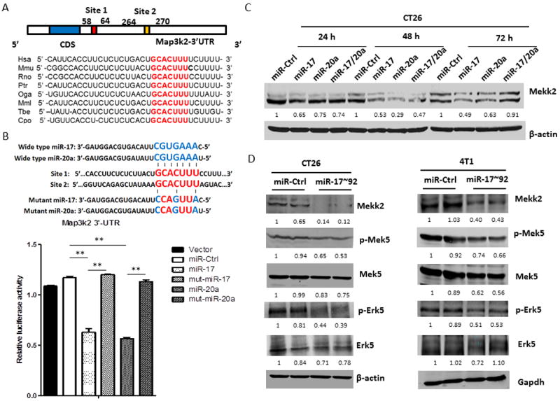
Figure 4. MiR-17/20a suppresses the expression of MHC class I via the Erk5 signal pathway by targeting Mekk2.

(A) Schematic representation of the mouse Map3k2 3’-UTR. MiR-17/20a complementary sites are indicated (vertical red lines).
(B) Schematic representation of mutant sites of miR-17 and miR-20a (upper panel). HEK293T cells were co-transfected with wild-type Map3k2 3’-UTR luciferase reporter plasmids, together with miR-17 mimic, miR-20a mimic or mutant miR-17, mutant miR-20a (Thermo scientific) as indicated. Renilla luciferase activity was measured 24 hours after transfection. Error bars represent standard deviation (±SD) (one-way ANOVA; ** p<0.01).
(C) Western blots showing expression of Mekk2 in CT26 cells after transient transfection with miR-Ctrl, miR-17, miR-20a or miR-17/20a for 24, 48 or 72 hours. β-actin was used as a loading control.
(D) Western blots showing expression of Mekk2, p-Mek5, Mek5, p-Erk5 and Erk5 in CT26/miR-Ctrl and CT26/miR-17~92 cell lines (left panel) or 4T1/miR-Ctrl and 4T1/miR-17~92 cell lines (right panel). β-actin or Gapdh was used as a loading control.
