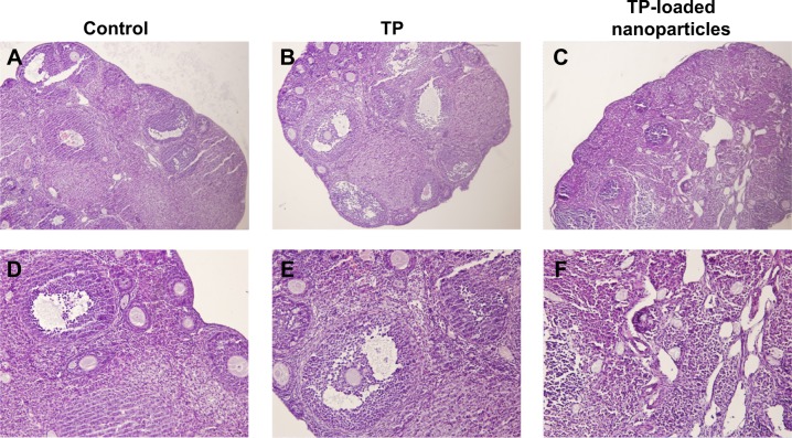
Figure 6.
The changes in ovarian histomorphology in the three groups.
Notes: (A–C): hematoxylin and eosin ×100; (D–F): hematoxylin and eosin ×200. Different stages of developing follicles and distinct corpus luteum were clearly observed in the control and TP groups. Follicle development was suppressed in the TP-loaded nanoparticle group, and few corpus lutea were observed. Ovarian tissue was seriously damaged, with overt ovarian fibrosis and vacuolar changes.
Abbreviation: TP, triptolide.

