
Figure 3. Cell structure of Chlamydomonas reinhardtii.
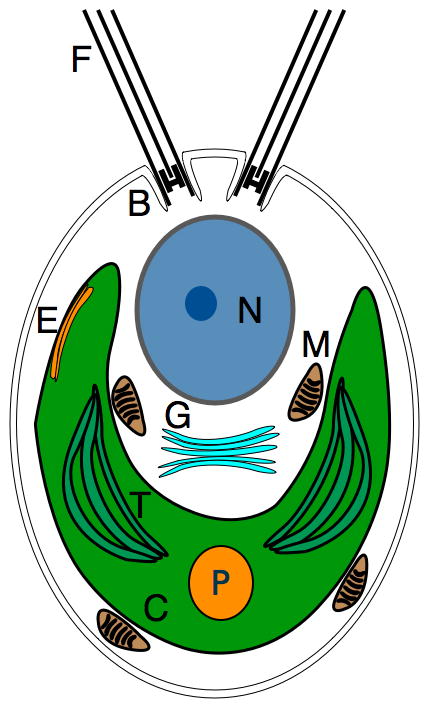
Schematic representation of the subcellular components of a typical Chlamydomonas cell; flagella (F), basal body (B), nucleus (N), eye spot (E), golgi apparatus (G), mitochondria (M), chloroplast (C), thylakoids (T), and the pyrenoid (P).